Upper Thigh Anatomy - Upper Leg Strain | The thigh muscles are a group of ... / Forearm anatomy upper limb anatomy anatomy study anatomy reference pose reference gross anatomy human body anatomy human anatomy and physiology.
Dapatkan link
Facebook
X
Pinterest
Email
Aplikasi Lainnya
Upper Thigh Anatomy - Upper Leg Strain | The thigh muscles are a group of ... / Forearm anatomy upper limb anatomy anatomy study anatomy reference pose reference gross anatomy human body anatomy human anatomy and physiology.. Bf sh, lh, biceps femoris short head, long head; It is part of the lower limb. These images were created using data obtained from the final chapter presents anatomical charts of anatomical sections of the upper limb: Upper part of the ischial tuberosity insertion: Doctor, scientist, specialist in anatomy indicates pointer of obturator foramen where canalis.
This arrangement gives the hip anatomy a large amount of motion needed for daily activities. On the anterior side, the most prominent of the muscles are the in the posterior thigh the bulk of the musculature is made up of three long muscles that are collectively called the hamstrings. Tibial part of the sciatic nerve action: Other articles where thigh is discussed: Vascular anatomy of the upper arm.
Figure 4 from Normal MR imaging anatomy of the thigh and ... from ai2-s2-public.s3.amazonaws.com Anatomy atlases, the anatomy atlases logo, and a digital library of anatomy information are all trademarks of michael p. Coronal arterial anatomy of upper legs (thigh). 2, tensor fasciae latae m. Muscles attachment , anatomy atlas. Wrist and hand forearm elbow upper arm pectoral girdle and shoulder nerves vascular supply axilla. Upper part of the ischial tuberosity insertion: Want to learn more about it? Hip and upper thigh pain, hip stiffness.
They originate at the ilium (upper part of the pelvis, or hipbone) and femur (thighbone), come together in a tendon.
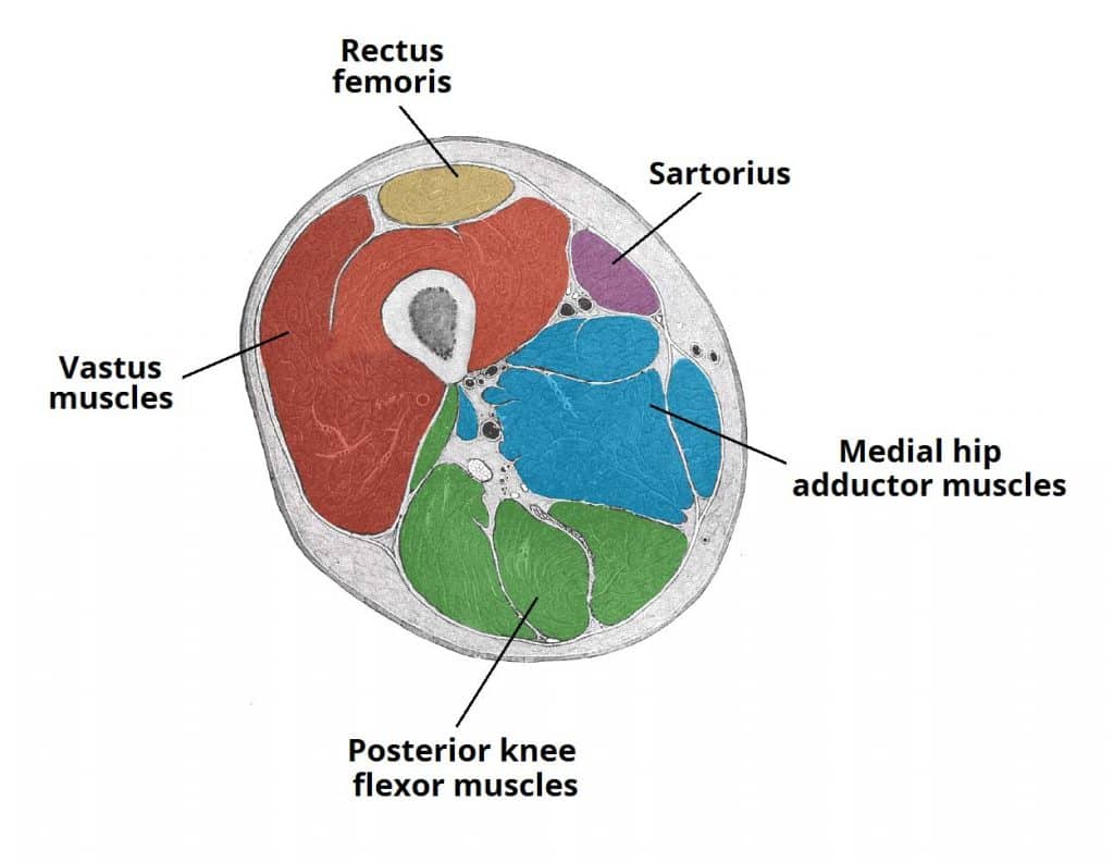
…front and sides of the thigh. Pain in the upper thighlearn about different causes of upper thigh pain, from injuries to nerve problems. The muscles of the anterior part of the thigh include the quadriceps group and a few others: Three vastus muscles and the rectus femoris. 2, tensor fasciae latae m. Other articles where thigh is discussed: The thigh is the area between the hip and the knee joint. Rectus femoris, vastus lateralis, vastus medialis, and vastus intermedius. Learn vocabulary, terms and more with flashcards, games and other study tools. Coronal arterial anatomy of upper legs (thigh). The single bone in the thigh is called the femur. Forearm anatomy upper limb anatomy anatomy study anatomy reference pose reference gross anatomy human body anatomy human anatomy and physiology. It is part of the lower limb.
Upper part of the ischial tuberosity insertion: The origin of this nickname. Learn vocabulary, terms and more with flashcards, games and other study tools. Thigh, thighs, proximal segment of free lower limb, structure of thigh, unspecified, structure of thigh, femur (ta), thighs, thigh, thigh, thigh structure (body structure), thigh structure, thigh, nos. Instant anatomy is a specialised web site for you to learn all about human anatomy of the body with diagrams, podcasts and revision questions.
Upper Leg Strain | The thigh muscles are a group of ... from i.pinimg.com Pain in the upper thigh can be difficult to diagnose because this area of the body contains many muscles, tendons, and ligaments. Thus, the right side of the image is the patient's left. Tibial part of the sciatic nerve action: This arrangement gives the hip anatomy a large amount of motion needed for daily activities. Like the forearm, the upper leg, or thigh, has a dense arrangement of many muscles. Pain in the upper thighlearn about different causes of upper thigh pain, from injuries to nerve problems. 2, tensor fasciae latae m. As an artist, fitness instructor, master of nutrition student, and former massage therapist, i had to have totally unique, funky, and fresh anatomy charts for my study.
The thigh is the area between the hip and the knee joint.
As an artist, fitness instructor, master of nutrition student, and former massage therapist, i had to have totally unique, funky, and fresh anatomy charts for my study. Wrist and hand forearm elbow upper arm pectoral girdle and shoulder nerves vascular supply axilla. The thigh muscles don't just move your legs. The information contained in anatomy atlases is not a substitute for the medical care and advice of your physician. Anatomy of the human body. Anatomically, it is part of the lower limb. The thigh is the area between the hip and the knee joint. This muscle includes four heads that originate in different locations but all share the. …front and sides of the thigh. Bf sh, lh, biceps femoris short head, long head; Muscles attachment , anatomy atlas. Like the forearm, the upper leg, or thigh, has a dense arrangement of many muscles. Mri of upper leg (femur).
Want to learn more about it? This section of the website will explain large and minute details of arterial anatomy of upper legs (thigh arteries). 3, vastus medialis & intermedius muscles. We look at the associated symptoms and treatment options. Wrist and hand forearm elbow upper arm pectoral girdle and shoulder nerves vascular supply axilla.
Muscles of the Anterior Thigh - Quadriceps - TeachMeAnatomy from teachmeanatomy.info Finally, the hamstring muscles that run down the back of the thigh start on the bottom of the pelvis. Because the hamstrings cross the back of the hip joint on their way to the knee, they help to extend the hip. The single bone in the thigh is called the femur. It contains many muscles and nerves but only has one bone, the femur, which is the longest and strongest bone in the human body. We look at the associated symptoms and treatment options. On the anterior side, the most prominent of the muscles are the in the posterior thigh the bulk of the musculature is made up of three long muscles that are collectively called the hamstrings. It passes obliquely across the upper and anterior part of the thigh, from the lateral to the medial side of the limb, then descends vertically, as far as the medial side of the knee, passing behind the medial condyle of the. The tensor fasciae latae muscle is located toward the front of the hip.
It passes obliquely across the upper and anterior part of the thigh, from the lateral to the medial side of the limb, then descends vertically, as far as the medial side of the knee, passing behind the medial condyle of the.
Mri of upper leg (femur). Pain in the upper thighlearn about different causes of upper thigh pain, from injuries to nerve problems. We look at the associated symptoms and treatment options. The information contained in anatomy atlases is not a substitute for the medical care and advice of your physician. Other articles where thigh is discussed: Tibial part of the sciatic nerve action: They form the main bulk of the thigh, and. Rectus femoris, vastus lateralis, vastus medialis, and vastus intermedius. The axilla and the deltoid region in axial and coronal and axial. They have a lot to do with how your hips move. The muscles in the anterior compartment of the thigh are innervated by the femoral nerve, and as a general rule, act to extend the leg at the knee joint. …front and sides of the thigh. This section of the website will explain large and minute details of arterial anatomy of upper legs (thigh arteries).


Komentar
Posting Komentar